Code
Our research is focused on the convergence of modern data science, high-performance computing, and materials science. In pursuit of this, we are developing new ways to extract rich, physically-meaningful information from complex data streams at high speed. We are also using artificial intelligence and machine learning to explore and harness multidimensional data streams more quickly and effectively. On this page, you will find code from our work on sparse data analytics, human-computer interaction, and electron microscopy data analysis.
Code is provided for research purposes under the terms of the respective repository licenses. No warranty of fitness for a particular purpose is expressed or implied.
Catalyst Vision: Quantifying Nanocatalyst Structure
and Evolution with Computer Vision
Developing active and durable nanocatalysts is essential for advanced energy technologies, including fuel cells and electrolyzers. The performance of these materials is directly linked to their nanoscale structure, such as particle size, shape, and distribution on a support material. However, quantifying these structural parameters and tracking how they evolve during operation is a significant challenge. This often requires tedious manual analysis of microscopy images, which is slow, subjective, and difficult to scale.
Catalyst Vision addresses this challenge using a human-in-the-loop, deep-learning-based workflow. Our software empowers users to train a powerful semantic segmentation model with only a few examples, enabling the rapid and automated analysis of large electron microscopy datasets. By accurately identifying and measuring features like nanoparticles and support materials, Catalyst Vision extracts the critical statistical information needed to build robust structure-property relationships. This workflow is designed to accelerate the development of predictive models for catalyst performance and degradation, paving the way for more resilient and efficient energy materials.
MORE INFORMATION
Chan, A.-L., Hayden, S., Harvey, S.P., Smeaton, M., Okrucky, C., Watt, J., Ulična, S., Spurgeon, S.R., Jungjohann, K., and S. Alia. “Mechanism-informed breakdown: understanding degradation by controlling voltage hold patterns in PEM water electrolyzers.” Preprint (2025). [DOI:10.2139/ssrn.5534899]
To download the code, visit: https://github.com/NREL/Catalyst-Vision
A look at the Catalyst Vision graphical user interface. Our human-in-the-loop, deep learning software enables rapid semantic segmentation of nanocatalyst features, providing the critical statistical data needed to link material structure to performance.
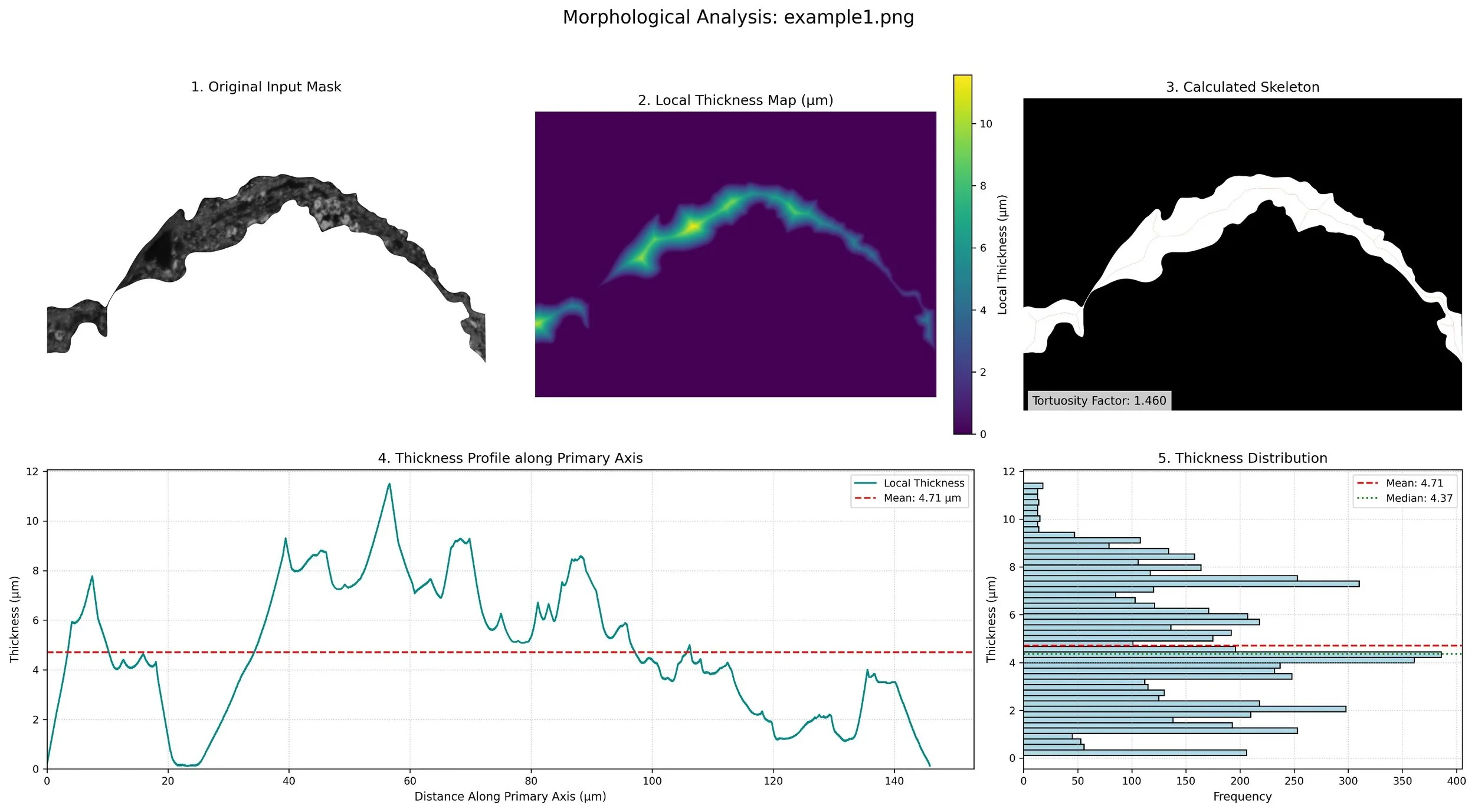
GraphEM: Extracting Order Descriptors Through Multi-Modal Graph Analytics
Describing order in materials is a critical step in achieving performance for applications such as energy storage, computing, and sensing. However, order is encoded across multiple data modalities, which are often challenging for humans to interrogate and interpret. Graph-based segmentation overcomes these challenges by representing each pixel in an image as a node in a graph, where the edges of the graph are weighted by the similarity between pixels. The graph is then partitioned into segments using a variety of algorithms. This approach is well-suited for the analysis of complex materials microstructures, particularly when multiple imaging modalities are used. We used a combination of high-angle annular dark-field (HAADF) scanning transmission electron microscopy (STEM) and energy dispersive X-ray spectroscopy (EDS) to characterize the microstructure of a complex material. The HAADF images provided information about the morphology of the material while the EDS data provided information about the chemical composition. By combining these two modalities, we were able to segment the images into different phases and identify the chemical composition of each phase. This information was then used to train a machine learning model to predict the properties of the material.
More Information
Ter-Petrosyan, A., Holden, M., Bilbrey, J.A., Akers, S., Doty, C., Yano, K.H., Wang, L., Paudel, R., Lang, E., Hattar, K., Comes, R.B., Du, Y., Matthews, B.E., and S.R. Spurgeon. “Revealing the evolution of order in materials microstructures using multi-modal computer vision.” Arxiv Preprint (2024). [DOI:10.48550/arXiv.2411.09896]
To download the code, visit: https://github.com/pnnl/GraphEM

Multi-modal analytics exhibits greater discriminating power to describe crystalline order.
EELSTM: Forecasting of In Situ EELS Data
Reward-based decision-making is directly linked to our ability to accurately forecast, or anticipate, changes in a system or process. Effective forecasting is essential for many disciplines and technologies we take for granted, ranging from meteorology to the power grid and from stock trading to logistics. The recent rise of autonomous vehicles, including automobiles, drones, and spacecraft, has been propelled by advanced forecasting models deployed on high performance computing platforms. Abundant low-cost computing and the proliferation of machine learning (ML) have enabled many new real-time forecasting approaches. When performed correctly, forecasting can save time, reduce cost, and guide scientific discovery by helping direct decision-making. We have developed deep learning-based approaches to forecasting of in situ imaging and electron energy loss spectroscopy data, allowing us to anticipate and respond to changes in materials and chemical systems for self-driving experimentation.
Students performing this research were supported by the UW Industry Capstone Program and the National Science Foundation.
More Information
Lewis, N., Jin, Y., Tang, X., Shah, V., Doty, C., Matthews, B.E., Akers, S. and S.R. Spurgeon. “Forecasting of in situ electron energy loss spectroscopy.” npj Computational Materials. 8 (2022): 252. DOI:10.1038/s41524-022-00940-2[Download PDF]
Fu, W., Spurgeon, S.R., Wang, C., Shao, Y., Wang, W. and A. Peles. “Deep-learning-based prediction of nanoparticle phase transitions during in situ transmission electron microscopy.” Arxiv Preprint. https://arxiv.org/abs/2205.11407
To download the code, visit: https://github.com/pnnl/EELSTM
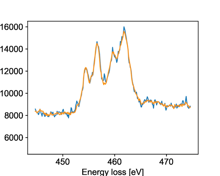
Raw (blue) and forecasted (golden) EELS data for Ti L edge EELS collected during in situ reduction.
pyCHIP: A Graphical User Interface for Few-Shot Machine Learning-Based Segmentation
The recent growth in data volumes produced by modern electron microscopes requires rapid, scalable, and flexible approaches to image segmentation and analysis. Few-shot machine learning, which can richly classify images from a handful of user-provided examples, is a promising route to high-throughput analysis. However, current command-line implementations of such approaches can be slow and unintuitive to use, lacking the real-time feedback necessary to perform effective classification. pyCHIP is a Python-based graphical user interface that enables end users to easily conduct and visualize the output of few-shot learning models. This interface is lightweight and can be hosted locally or on the web, providing the opportunity to reproducibly conduct, share, and crowd-source few-shot analyses.
Students performing this research were supported by the UW-DIRECT Data Science Capstone Program and the National Science Foundation.
More Information
Doty, C., Gallagher, S., Cui, W., Chen, W., Bhushan, S., Oostrom, M., Akers, S., and S.R. Spurgeon. “Design of a graphical user interface for few-shot machine learning-based classification of electron microscopy data.” Computational Materials Science. 203.15 (2021): 111121. DOI:10.1016/j.commatsci.2021.111121[Download PDF]
Akers, S., Kautz, E., Trevino-Gavito, A., Olszta, M., Matthews, B., Wang, L., Du, Y., and S.R. Spurgeon. “Rapid and flexible segmentation of electron microscopy data using few-shot machine learning.“ npj Computational Materials. 7 (2021): 187. DOI:10.1038/s41524-021-00652-z[Download PDF]
To download the code, visit: https://github.com/pnnl/pychip_gui
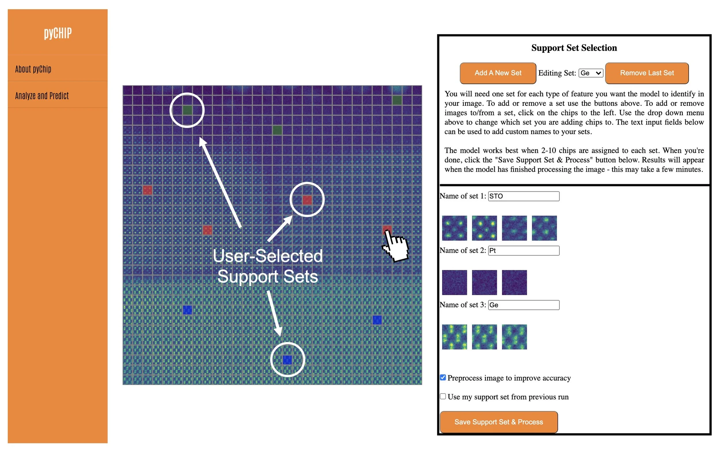
Demonstration of pyCHIP functionality and few-shot-based TEM image classification.
TEMWizard: A GUI for Atomap Image Analysis
Extraction of quantitative crystallographic information from atomic-resolution transmission electron microscopy (TEM) images is challenging, since data are often noisy and artifacted. The Atomap package permits fitting and analysis of key features in atomic-resolution data, but its command-line format does not lend itself to facile and dynamic exploration of image data. TEMWizard provides an easy-to-use graphical user interface (GUI) for Atomap, allowing the user to provide correct inputs and visualize lattice displacements for analysis of phenomena such as octahedral rotations or bond distortions.
More Information
Wang, L., Zhao, J., Kuo, C-T., Matthews, B.E., Oostrom, M.T., Spurgeon, S.R., Bowden, M.E., Lee, S-J., Lee, J-S., Guo, E-J., Wang, J., Chambers, S.A., and Y. Du. “Synthesis and electronic properties of epitaxial SrNiO3/SrTiO3 superlattices.” Physical Review Materials. 6 (2022): 075006. DOI:10.1103/PhysRevMaterials.6.075006
To download the code, visit: https://github.com/pnnl/temwizard
To learn more about Atomap, visit: https://atomap.org/

Demonstration of real-time visualization of lattice plane spacings and distortions using TEMWizard.
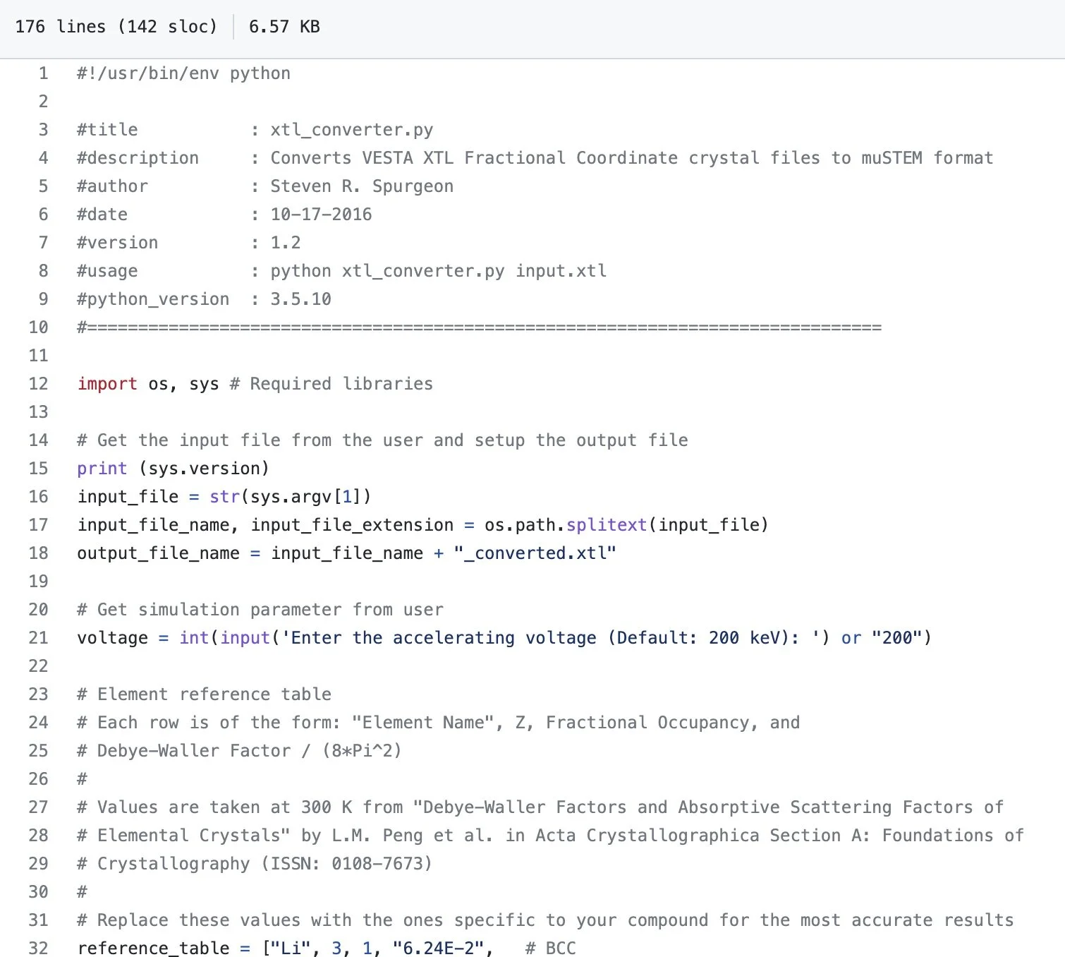
xtl-converter: Data Preparation for µSTEM TEM Image Simulation
Image simulation is an important part of quantitative analysis of electron microscopy data. The µSTEM package (https://github.com/HamishGBrown/MuSTEM) allows for rigorous simulation of images and ionization maps to aid in this process. However, the preparation of data from crystal files requires extensive reformatting and can be time-consuming. The xtl-converter code aids in this process by reformatting VESTA fractional coordinates (*.xtl) files into the proper configuration.
More Information
Spurgeon, S.R., Du, Y., and S.A. Chambers. “Measurement error in atomic-scale scanning transmission electron microscopy–energy-dispersive X-ray spectroscopy (STEM-EDS) mapping of a model oxide interface.” Microscopy and Microanalysis. 23.3 (2017): 513–517. DOI:10.1017/S1431927617000368[Download PDF]
To download the code, visit: https://github.com/stevenspurgeon/xtl-converter
Conversion of crystallographic file formats for easy handling using the µSTEM simulation package.